Confirming placement of endotracheal tube: Monitoring techniques endotracheal tube
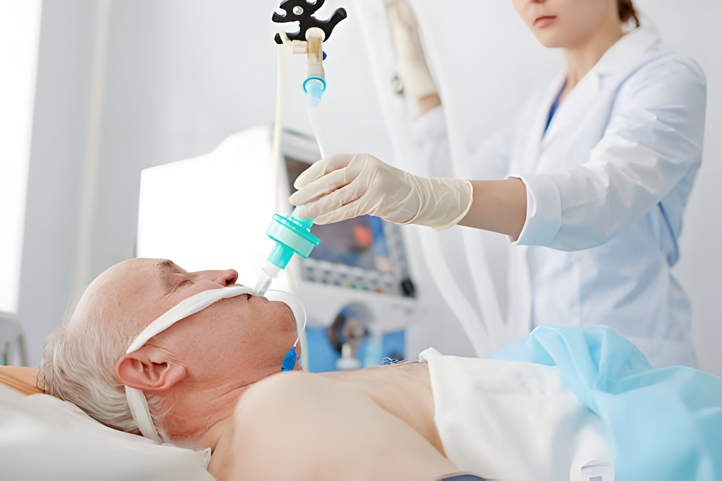
The most critical aspect of airway management during medical procedures is ensuring that the placement of an endotracheal tube (ETT) is accurate. This is because incorrect placement can lead to severe complications like hypoxemia and aspiration. So, several monitoring techniques are employed to confirm the proper positioning of the ETT within the trachea.
By understanding these techniques, healthcare professionals can improve patient outcomes during airway management procedures. Let us explore these monitoring techniques, and procedures further.
What are the primary techniques used for confirming the placement of an endotracheal tube (ETT)?
The key techniques used for confirming the placement of an endotracheal tube include the following.
- Capnography
- Auscultation
- Chest rise and fall
- Chest X Ray
- Ultrasound
- Colorimetric devices
- Bilateral breath sounds
How does capnography aid in verifying ETT placement?
Capnography aids in confirming the placement of an endotracheal tube (ETT) by measuring the concentration of exhaled carbon dioxide (CO2). Here’s how:
- Detects the presence of exhaled CO2, indicating ventilation is occurring.
- Provides immediate confirmation of tracheal placement by detecting CO2 in exhaled breath.
- Enables continuous monitoring of CO2 levels, ensuring ongoing confirmation of ETT position.
- Allows for quick detection of accidental extubation or displacement of the ETT.
- Differentiates between tracheal and esophageal intubation based on CO2 levels.
- Helps identify mainstem bronchus intubation by detecting unequal CO2 levels in each lung.
- Facilitates adjustment of ETT depth based on CO2 waveform characteristics.
- Helps assess the effectiveness of CPR by monitoring changes in CO2 levels during resuscitation.
Read more: ECG Waves Explained: A Beginner’s Guide
What role does auscultation play in confirming ETT placement?
Auscultation helps in confirming endotracheal tube (ETT) placement by assessing breath sounds. Here’s how it aids in confirmation:
| Auscultation Role |
Description |
| Bilateral breath sounds |
Ensures equal breath sounds over lung fields, indicating proper ETT placement. |
| Absence of gastric sounds |
Confirms no sounds over the epigastrium, indicating tracheal rather than esophageal placement. |
| Axillary breath sounds |
Detects potential mainstem bronchus intubation, requiring reassessment. |
| Monitoring during ventilation |
Confirms ETT patency and proper ventilation during manual ventilation efforts. |
| CPR assessment |
Evaluate resuscitation effectiveness and detect changes in lung sounds. |
| Transport monitoring |
Allows ETT placement assessment and immediate detection of displacement. |
| Postintubation verification |
Essential for confirming proper ETT placement and ensuring patient safety. |
What are the key sounds that indicate proper positioning?
Here are the key sounds indicating correct placement:
- Bilateral equal breath sounds over lung fields.
- Absence of gastric sounds over the epigastrium.
- No breath sounds in the axillae, indicating avoidance of mainstem bronchus.
- Continuous, audible breath sounds during bag valve mask ventilation.
- Consistent breath sounds during cardiopulmonary resuscitation.
- Persistent breath sounds during patient transport.
- Verified absence of air leak around ETT cuff.
How do you perform chest auscultation to confirm ETT placement?
Chest auscultation is a vital method for confirming the placement of an endotracheal tube (ETT). Here’s how it’s performed:
Positioning:
Ensure the patient is in a supine position with the head slightly elevated.
Stethoscope Placement:
Place the diaphragm of the stethoscope over the lung fields bilaterally.
Breath Sounds:
Listen for equal and symmetrical breath sounds over both lung fields.
Gastric Sounds:
Check for the absence of gastric sounds in the epigastrium.
Axillary Region:
Assess for breath sounds in the axillae to detect potential mainstem bronchus intubation.
Manual Ventilation:
During bag-valve-mask ventilation, keep monitoring for consistent and audible breath sounds.
CPR Assessment:
Evaluate breath sounds during cardiopulmonary resuscitation for changes indicative of ETT displacement.
Transport Monitoring:
Maintain auscultation during patient transport to ensure continuous confirmation of ETT placement.
What are the benefits and drawbacks of using chest X-rays to verify ETT placement?
Chest X-rays give us a proper confirmation of endotracheal tube (ETT) placement but come with its benefits and drawbacks. The benefits and drawbacks include the following;
Benefits:
- Visual confirmation of ETT position.
- Assessment of complications.
- A permanent record for documentation.
Drawbacks:
- Delay in confirmation.
- Radiation exposure.
- Resource intensive.
- Costly.
- Interpretation variability.
What is the significance of observing chest rise and fall during manual ventilation in confirming ETT placement?
Observing chest rise and fall during manual ventilation is significant for confirming endotracheal tube (ETT) placement. The symmetrical chest expansion shows that the ETT is correctly positioned within the trachea. This ensures that there is adequate airflow to both lungs. Inconsistent chest movement is a sign of ETT displacement. This needs quick intervention to prevent hypoventilation or hypoxia.
How does the use of ultrasound contribute to verifying ETT placement?
Ultrasound offers a dynamic method for confirming endotracheal tube (ETT) placement. Here’s how it contributes:
Real Time Visualization:
Provides immediate visualization of ETT within the trachea.
Direct Imaging:
Allows direct visualization of ETT position relative to surrounding structures.
Reduced Radiation:
Eliminates the need for ionizing radiation exposure associated with chest X-rays.
Portable and Accessible:
Can be performed bedside, enabling rapid assessment in many clinical settings.
Continuous Monitoring:
Allows monitoring of ETT placement during procedures and patient transport.
Read more: Things You Need to Know About PEA
Conclusion
Making sure the endotracheal tube is in the right place is crucial for keeping patients safe during medical procedures. There are different ways to check this, like listening to breath sounds, using special machines, taking X-rays, and using ultrasound. Each method has its advantages and demerits, but using them together to achieve the best patient outcomes is the key. By using these techniques, healthcare professionals can help save patients’ lives when they need support in breathing.
Resources:
PALS CERTIFICATION
Author
PALS Certification is a trusted provider of online life support training, offering PALS, BLS, and ACLS certification and renewal courses. Our flexible training programs follow industry guidelines, offer self-paced learning and instant certification, ensuring providers stay compliant, advance their credentials, and deliver high-quality patient care.